
In Wei Sun's Biofabrication Lab in the College of Engineering, researchers have figured out a way to print 3-D blocks of living stem cells.
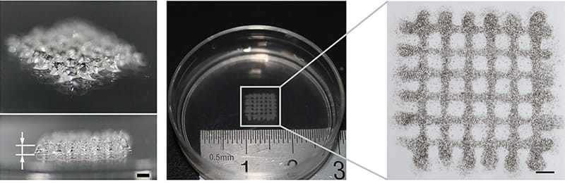
Using a special designed extrusion printer he created to squeeze out a mixture of hydrogel and stem cells, Wei Sun, PhD, Albert Soffa chair professor in Drexel’s College of Engineering, is making strides toward rapid prototyping the building blocks of life. His process, which was recently published in Biofabrication gives scientists a head start at growing living three-dimensional tissues and could one day be used to create micro-organs for research purposes.
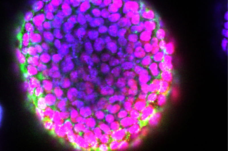
“We are able to apply a 3-D printing method to grow embryoid body (EB) in a controlled manner to producing highly uniform ‘blocks’ of embryonic stem cells,” Sun said. “These EB ‘blocks’ are capable of differentiating to other cell types and could be used as the ‘lego bricks’ to build tissue constructs, larger structures of tissues, and potentially even micro-organs.”
Sun’s method, along with the specialized bio-printer used to carry it out, have been his primary research thrusts over the last decade. The bio-printer, which was originally created with the goal of printing bone and tissue scaffolds, has also been modified to print living cancer tumors. It does so by extruding the cells in a temperature-controlled hydrogel mixture and depositing a three-dimensional grid-like structure, which not only keeps them viable, but helps them grow and multiply.
“I think we’ve produced a 3-D microenvironment which is more like that found in vivo for growing embryoid body, which explains the higher levels of cell proliferation,” said Sun, who co-authored the research with colleagues from Tsinghua University in Beijing. “There is still a long way to go from a varying sized EB to a regenerated organ, but our work provides a promising tool to facilitate this development.”
The benefit of being able to carefully manipulate the environment that exists in a “block” of stem cells is that it gives researchers a controlled platform from which to study how and why the cells differentiate into precursors of organs—called embryoid bodies. Down the road, this could mean that scientists will be able to more efficiently grow organs in a lab by printing and growing the organ’s corresponding embryoid body.
While this could eventually impact regenerative medicine, currently, the more approachable application is to rapidly grow tissues for drug screening studies. Printed blocks of stem cells grow more quickly and develop in a more natural way than those cultured in a petri dish, because they are surrounded, on all sides, by other cells—the same way they would be found in the body.
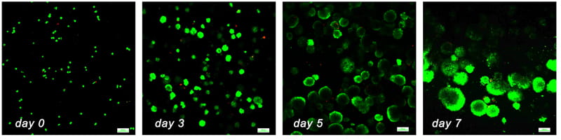
It is also significant to note that after printing, the stem cells not only thrive, but also retain their ability to differentiate— to grow into a variety of organs and tissues. It has been well documented that the size of the embryoid body is linked to gene expression and cell fate, so being able to control the size of the printed stem cell block equates to controlling which tissues and micro-organs are created. Studying and replicating this variability is the next research thrust for Sun’s Biofabrication Laboratory.
“Being able to produce varying sized embryoid bodies by tuning the 3-D printing process, materials and structural parameters, one can potentially bio-fabricate different cell types and potentially grow micro-organs from a printed 3-D biological model,” Sun said. “We are not directly going to printing an organ, but we can print an in vitro 3-D biological model which could lead to growing different size embryoid bodies, different types of cells, and, ultimately, to growing a regenerated organ. This will be a significant advance for stem cell research and for regenerative medicine.”
Sun’s research was supported in part by a grant from the Drexel-SARI Research Center in Shanghai.