Facilities & Instrumentation
Biosensor Technology
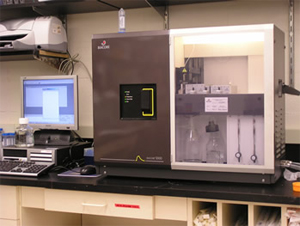
The Chaiken lab in the Department of Biochemistry operates a biosensor facility, featuring a Biacore 3000 surface plasmon resonance instrument, used for detecting interactions between biological macromolecules.
Back to Top
Capillary Electrophoresis
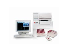
Our Beckman Coulter P/ACE MDQ capillary electrophoresis system provides a versatile, efficient, and sensitive technique for the separation of biomolecules. The option exists to pipe the output from the CE system directly to our electrospray mass spectrometer.
Back to Top
Fermentation
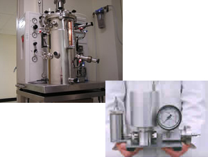
Facilities for large-scale growth of bacterial and yeast cultures include eight temperature-controlled floor shakers, a walk-in 37°C chamber, and a 20-liter New Brunswick BioFlow IV fermenter (with a dedicated CEPA continuous flow centrifuge for harvesting cell pellets). An Avestin Emulsiflex C-5 is available for rapid and efficient breakage of cells for protein purification.
Back to Top
Fluorescence Microscopy
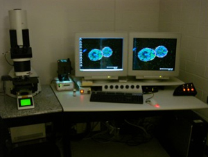
The Reginato lab operates the confocal facility featuring the Leica Laser Scanning Spectral Confocal Microscope TCS SP2 AOBS advanced image acquisition and analysis system. This system includes a UV laser, as well as standard argon, and helium neon lasers that can simultaneously image four different fluorophores. In addition, the microscope includes objectives of 20x, 40x, 63x, and 100x magnification.
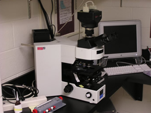
In addition, the Department has an Olympus IX71 inverted epifluorescence microscope with MetaMorph/MetaFluor Imaging System, CoolSnap HQ Digital CCD camera, and DG4 Galvo-wavelength switcher as well as an Olympus AX70 Compound epifluorescence microscope with Spot RT Slider camera.
Back to Top
Protein Interaction Array System
The ProteOn™ XPR36 protein interaction array system is an SPR optical biosensor that provides real-time data on the affinity, specificity, and interaction kinetics of protein interactions. Using XPR technology, a unique approach to multiplexing, this system generates a 6 x 6 interaction array for the simultaneous analysis of up to six ligands with up to six analytes. The ProteOn XPR36 system increases the throughput, flexibility, and versatility of experiment design, enabling the completion of more experiments in less time:
- Analyze up to 36 different protein interactions in a single run, on a single chip
- Perform a complete kinetic analysis in a single run
- Measure a variety of experimental conditions simultaneously in parallel
- Screen multiple panels of analytes
Back to Top
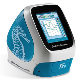
Seahorse Analyzer XFp Analyzer
The department has a Seahorse Analyzer XFp Analyzer. This instrument measures cellular metabolism in live cells in real time, via the measurement of extracellular flux (oxygen consumption rate and extracellular acidification rate) to provide insight into mitochondrial function and glycolytic activity. XF technology can be used to determine which pathway is predominant (metabolic phenotype), as well as which substrates are being used for energy production. Glucose oxidation, fatty acid oxidation, and aerobic glycolysis rates can be readily measured under multiple conditions.
Back to Top
Spectroscopy
The Jorns laboratory operates a Hi-Tech Scientific Stopped-Flow System (SF-61DX2) that is equipped for anaerobic single- or double-mixing experiments with multiple detection options, including single wavelength and diode array absorbance modes.
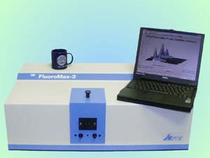
Shared facilities with the Department feature a Jasco J810 CD spectrometer and a Fluoromax-3 Fluorimeter (Spex).
Back to Top
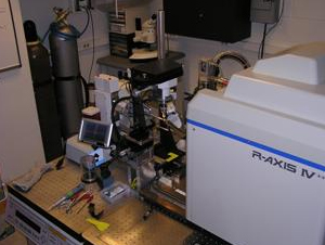
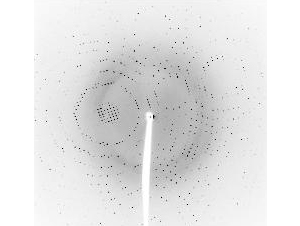
Structural Biology Center
Full understanding of molecules requires information about both structure and function. Structural analysis utilizes chemical and/or physical tools to unravel the links between the three-dimensional structure of a biologically important molecule (protein, DNA, or RNA) and its function.
We have been flooded with new information about the human genome. Translation of this information into structural information should rapidly lead to insights regarding function, which in turn are essential to the desired ultimate clinical applications. Now that we know that the precise regulation of cell function and organismic organization is accomplished by only 35,000 genes, it is clear that combinatorial processes must underlie the events controlling normal development and function and that aberrations in those control mechanisms are the root of disease. The molecular bases of these combinatorial events lie in protein-protein interactions; to properly understand them, it is essential to define these protein interactions at a three-dimensional level.
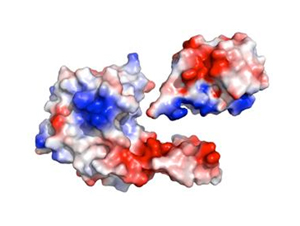
The Structural Biology Laboratories are housed within the Department of Biochemistry & Molecular Biology and are under the direction of Dr. Patrick Loll. The goal for the laboratories is to utilize a multifaceted approach to understanding structure/function relationships, in order to provide the structural information essential for understanding biologically important processes at the molecular level.
Back to Top
Miscellaneous
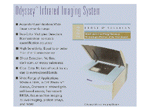
The department has acquired an Odyssey infrared imaging system from LI-COR Biosciences. This is a powerful technique that can enhance the sensitivity of western blots as well as a variety of other assays.
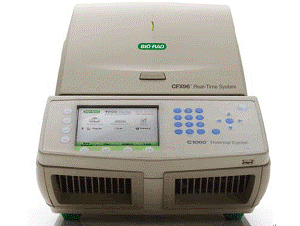
The Bio-rad CFX96™ optical reaction module converts a C1000™ thermal cycler into a powerful and precise real-time PCR detection system. This six-channel real-time PCR system comb has precise thermal control and delivers sensitive, reliable detection. Features include:
- Range of excitation/emission wavelengths: 450–730 nm
- Data collection from all wells during data acquisition
- Multiple data acquisition modes, including a 1-color fast scan for SYBR® Green users
- Thermal gradient feature to optimize reactions in a single run with ramp rate of 5°C/sec
- CFX Manager™ software with advanced analysis tools for performing normalized gene expression

Also available in the Department of Biochemistry & Molecular Biology:
- Two Beckman liquid scintillation counters
- Various high pressure liquid chromatography systems, including a Varian preparative-scale HPLC with automated sample changer, Multiple Beckman System Gold HPLCs, GE Akta FPLCs, and an Eksigent NanoLC
- Virtis Benchtop 4.0 ZL Lyophilizer
- MicroCal VP-ITC isothermal titration calorimeter
- Typhoon FLA 700 biomolecular imaging system
Back to Top