Optical Brain Imaging
1.1. Basic Theory of fNIR Imaging
All biological tissue is, to differing extents, permeable to electromagnetic (EM) radiation of different frequencies and intensities. This can also be considered as permeability of biological tissue to photons of different energy levels. This principle constitutes the basis of all imaging techniques that rely on transmission/scattering characteristics electromagnetic radiation, such as x-ray, Computed Axial Tomography (CAT), and photons for Near Infrared imaging. From the principles of spectroscopy, it is also known that different molecules absorb different wavelengths of EM radiation (which is synonymously referred to as light at smaller wavelengths) to different degrees, and likewise scatter that radiation to different degrees. In functional Near Infrared (fNIR) imaging, the molecule of concern is the hemoglobin molecule, which is the oxygen carrier for red blood cells. Since oxygenated hemoglobin, also referred to as oxy-hemoglobin or HbO2, absorbs light at a slightly different portion of the NIR spectrum than the deoxygenated hemoglobin molecules (usually referred to only as hemoglobin or Hb), it is possible to detect the relative concentrations of the two molecules using optical spectroscopic methods.
Photons emanating from any light source follow a characteristic path through the target tissue back to a detector that lies on the same approximate plane as the source (see Figure 1 below). While the light is severely attenuated due to the scattering and absorption processes, it is nonetheless encoded with the spectroscopic signatures of the molecules encountered en route to the detector. By carefully choosing the wavelengths that are produced by the source, it is possible to detect the relative concentrations of Hb and HbO2 in the target tissue. By comparing these levels to those obtained when the tissue is in its “baseline” state, and using some basic knowledge about “interesting” conditions for the tissue, it is possible to draw conclusions from systematic changes observed in these levels.
As a more concrete example, one may consider fNIR imaging of breast tissue. Here, the baseline condition of the breast tissue corresponds to its normal, healthy state. The interesting condition is the cancerous tissue. Since it is known that such tissue grows uncontrollably, the tissue must metabolize more than the baseline tissue. Therefore, to scan for tumors, one looks for concentrated areas of unusually high respiration levels.
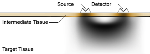
Figure 1. Simulated photon diffusion path through target tissue from source to detector. This simulation shows the photon path density, not the overall transmission level.
At the heart of the calculations used with fNIR imaging is the Beer–Lambert Law, which defines a linear relationship between absorption of EM radiation and the concentration of the target absorptive material in a given medium. The full law includes factors accounting for the absorption coefficient (wavelength dependent) and path length. Though originally defined for transmittance, it can be shown to be effective for diffusion/scattering as well. The measurements are initially taken in terms of received photon concentration, which can loosely be termed the “received power.” For biological fNIR imaging use, baseline data is first gathered. This reading is considered the “transmitter power.” Given these values, we can write the law as:

Since the law itself is linear, the total absorbance of any species is the sum of the absorbances of that species for each wavelength. Using these properties of the law, it is possible to take the received photon densities to calculate the Hb and HbO2 levels in the target medium in relation to the levels at baseline. This is a point that bears reiterating. The levels of Hb and HbO2 measured are relative to the baseline only. It is not possible to arrive at absolute values of concentration using fNIR imaging on living samples. Once the Hb and HbO2 levels are computed, they are used to calculate the levels of oxygenation (in mM), and values that may be approximately treated as percent changes in blood volume. The formulae are as follows:
